AI Glaucoma Detection: How Cup-to-Disc Ratio Analysis Enables Early Glaucoma Screening
Glaucoma screening is the attempt to find optic nerve damage before the patient notices any loss of vision. That is harder than it sounds, because glaucoma…
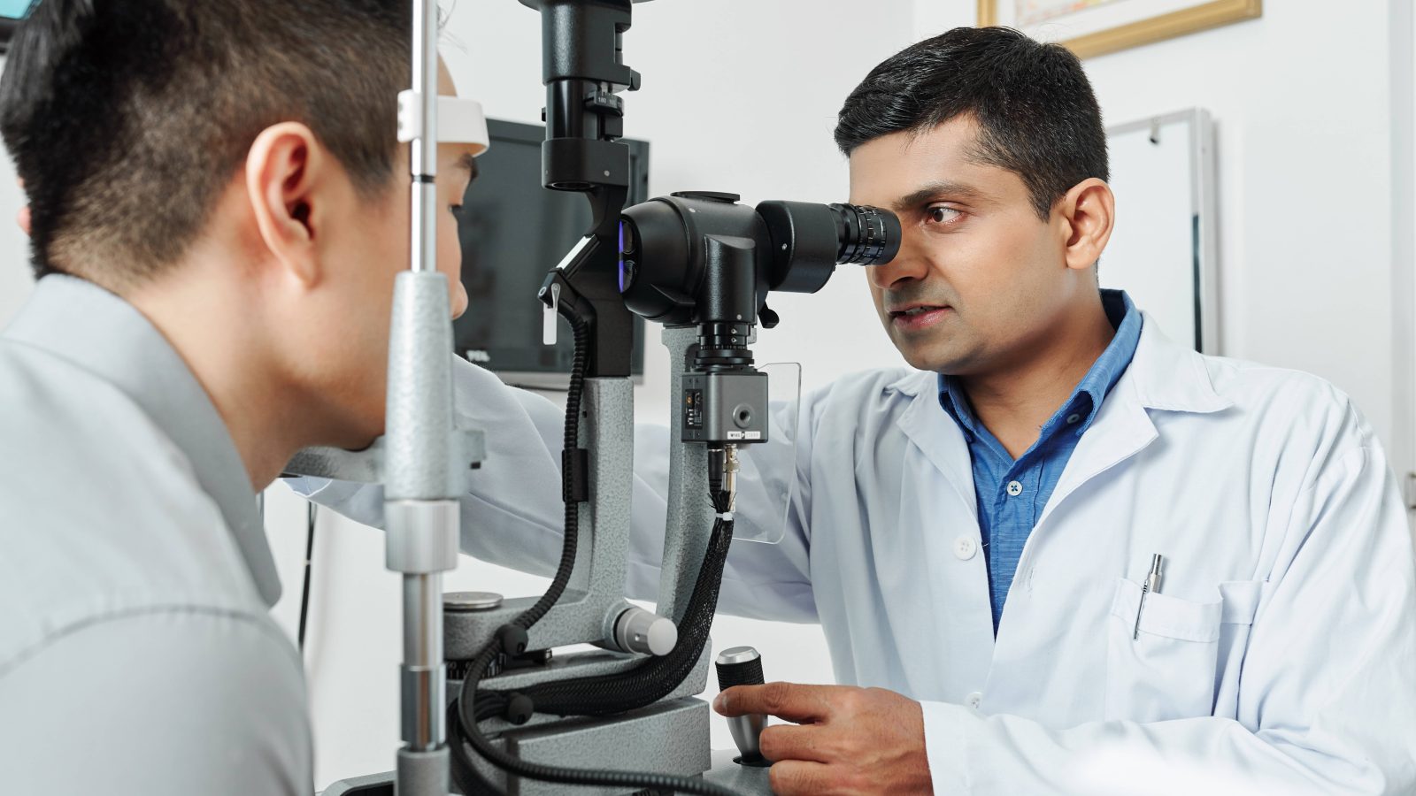
Diabetic retinopathy screening is the routine examination of the retina in people with diabetes to detect vascular damage before it threatens sight. The need in India is large. The ICMR-INDIAB study, published in The Lancet Diabetes and Endocrinology in 2023, estimated that 101 million Indians live with diabetes, and population studies place the prevalence of diabetic retinopathy at roughly 12 to 17 percent of that group. Guidelines recommend a retinal examination at least once a year for most people with diabetes, yet the number of ophthalmologists is far smaller than the number of eyes that need examining.
Deep learning changes the arithmetic. A trained algorithm can read a retinal photograph in seconds, assign a severity grade and flag which patients need to see an ophthalmologist. This article explains how AI diabetic retinopathy detection works from a single image, what the published evidence shows, and where the limits lie.
A fundus image is a colour photograph of the back of the eye that shows the optic disc, the macula and the retinal blood vessels. The lesions that define diabetic retinopathy are all visible on this photograph. They include microaneurysms, dot and blot haemorrhages, hard exudates, cotton wool spots, venous beading and new vessels. Fundus image analysis, whether by a human grader or an algorithm, is the search for these features and an assessment of their extent.
A non-mydriatic fundus camera captures a 45-degree field of the retina through an undilated pupil. The camera aligns on the eye using infrared light, which does not constrict the pupil, and then fires a brief visible flash to capture the image. Several current devices can image through a pupil of about 2 mm. Because no dilating drops are needed, there is no waiting period and no blurred vision afterwards, so screening can take place in a diabetes clinic, a primary health centre or a community camp rather than only in an eye hospital. Dilation is still used when the undilated image is too dark or too small to grade.
Grading follows the International Clinical Diabetic Retinopathy (ICDR) severity scale, a consensus classification published by Wilkinson and colleagues in Ophthalmology in 2003. It simplifies the 13-level ETDRS research scale into five clinical stages:
Diabetic macular oedema (DME) is graded separately as apparently present or apparently absent, because it can occur at any level of retinopathy.
Referable diabetic retinopathy is defined in the main validation studies as moderate NPDR or worse, referable DME, or both. This is the line that matters for screening. Patients below it can return for routine screening at the next interval. Patients at or above it need an ophthalmologist. Most AI systems are trained and validated against this threshold rather than asked to assign every one of the five grades with equal confidence.
The models used for AI diabetic retinopathy detection are convolutional neural networks. They are not programmed with rules about lesions. Instead, the network is trained on a large set of fundus photographs that ophthalmologists have already graded on the ICDR scale, and it learns which pixel patterns predict each grade. In the 2016 JAMA study by Gulshan and colleagues, the training set was 128,175 images and the labels came from a panel of 54 US-licensed ophthalmologists and senior trainees.
In use, the process runs in four steps.
The whole sequence takes seconds and produces a result on the spot, without a specialist present.
Three studies anchor the field.
Gulshan et al., JAMA, 2016. The algorithm was tested on two independent sets, EyePACS-1 (9,963 images) and Messidor-2 (1,748 images). At the high-sensitivity operating point, it reached 97.5 percent sensitivity and 93.4 percent specificity on EyePACS-1, and 96.1 percent sensitivity and 93.9 percent specificity on Messidor-2, for referable diabetic retinopathy. The reference standard was the majority decision of a panel of ophthalmologists.
Abràmoff et al, npj Digital Medicine, 2018. This was the pivotal trial behind IDx-DR, the first autonomous AI diagnostic system authorised by the US FDA, in April 2018. It enrolled 900 adults with diabetes and no prior retinopathy diagnosis at 10 primary care sites. Against a reading-centre standard, the system correctly identified 173 of 198 participants with more than mild diabetic retinopathy, a sensitivity of 87 percent. The images were taken by primary care staff, not eye specialists.
Gulshan et al., JAMA Ophthalmology, 2019. A prospective study of 3,049 patients with diabetes at Aravind Eye Hospital and Sankara Nethralaya in South India. The automated system reached 88.9 percent sensitivity and 92.2 percent specificity at Aravind, and 92.1 percent sensitivity and 95.2 percent specificity at Sankara Nethralaya. Manual grading by individual graders at the same sites ranged from 73.4 to 89.8 percent sensitivity. The authors concluded that the model generalised to an Indian population.
On the strength of this evidence, the American Diabetes Association has included FDA-authorised AI systems that detect more than mild retinopathy as an alternative to traditional screening in its Standards of Care since 2020, provided that referral pathways are in place.
AI screening addresses one step in the care pathway, which is image interpretation at the point of first contact. It moves that step into the diabetes clinic or primary care setting. It does not replace the ophthalmologist. A patient flagged as referable still needs a dilated examination, and often optical coherence tomography to confirm macular oedema, before any treatment decision is made.
Four limits are worth stating plainly.
The algorithm also looks only for diabetic retinopathy unless it has been trained for other conditions. Glaucoma, age-related macular degeneration and hypertensive changes may go unreported.
Diabetic retinopathy screening works because the disease announces itself on a photograph long before it affects vision. A non-mydriatic fundus camera makes that photograph easy to obtain in any clinic, the ICDR severity scale gives a shared language for grading it, and deep learning now reads it to the referable threshold with sensitivity and specificity comparable to trained human graders. The evidence from India specifically, where the model performed as well as or better than manual grading at two large eye hospitals, supports using this approach to reach the patients who are currently not screened at all.
Forus Health AI builds on this foundation with FH-POISE (Precision Ocular Intelligence for Systemic and Eye Health), its retinal image analysis platform. In February 2026, Forus Health received approval from the Central Drugs Standard Control Organisation (CDSCO) for FH-POISE DRT, a deep learning tool that assesses retinal images and predicts diabetic retinopathy severity. It is designed for use at the point of care in general hospitals, diabetes clinics, community health centres and outreach programmes, and it complements the 3nethra range of non-mydriatic fundus cameras that Forus Health manufactures. The aim is the one described throughout this article, which is to embed reliable screening into routine diabetes care so that referable disease is found early and specialist time is spent where it is needed.
Sources
Glaucoma screening is the attempt to find optic nerve damage before the patient notices any loss of vision. That is harder than it sounds, because glaucoma…